THE X-RAY TUBE
Components, operation and support systems in diagnostic radiology
ESSENTIAL SYSTEM COMPONENT
The X-ray tube is a component of the X-ray imaging system generally unknown to the radiology technician. It is located inside a protective housing and is, therefore, inaccessible during normal use. It is the heart of any radiology equipment.
MAIN COMPONENTS
Its components are considered separately, but it should be clear that there are two important parts: the cathode and the anode. Each of them is an electrode and any tube with two electrodes is a diode. An X-ray tube is a special type of diode.
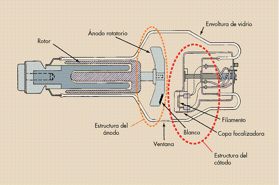
The internal structure of an X-ray tube consists of three main parts: the supporting structure, the protective housing and the metal or glass envelope. The internal structures of the X-ray tube are the anode and cathode.
CATHODE
Negative electrode that emits electrons through thermionic effect. Contains the tungsten filament that heats up to release electrons.
ANODE
Positive electrode (target) where electrons impact to produce X-rays. Can be stationary or rotating to better dissipate heat.
ENVELOPE
Pyrex glass or metal container that maintains internal vacuum. Provides electrical insulation and contains the beam output window.
WINDOW
Thin area (≈5 cm²) of the envelope that allows the useful X-ray beam to exit with minimal absorption. Generally made of beryllium in modern equipment.
TUBE SERVICE LIFE
With proper use, X-ray tubes used in general radiography should last many years. X-ray tubes used in computed tomography (CT) and interventional radiology generally have a much shorter life due to higher technical demands.
General Radiography
Extended service life (years)
Computed Tomography
Reduced service life (high demand)
Interventional Radiology
Shorter service life (intensive use)
EXTERNAL COMPONENTS AND SUPPORT SYSTEMS
The X-ray tube and housing are quite heavy. Therefore they require an assistance mechanism so the radiology technician can position them properly.
Ceiling Support System
Most common. Two perpendicular sets of ceiling rails allow longitudinal and transverse movement.
Floor-Ceiling System
Single column with rollers on ceiling and floor. The tube moves up and down the column when it rotates.
C-Arm
Used in interventional radiology. C-shaped mount on ceiling with great positioning flexibility.
SAFETY WARNING
Some X-ray tubes with ceiling support systems have a simple control that overrides the locks, allowing the tube to move freely. This override should only be used for small adjustments and should not be used to move the tube more than a meter, as shoulder and arm injuries may occur.
PROTECTIVE HOUSING
Isotropic radiation: When X-rays are produced, they are emitted isotropically, that is, with the same intensity in all directions. Only those X-rays emitted through the special section of the tube called the window are used.
X-rays that escape through the protective housing are called leakage radiation; this radiation does not contribute to diagnostic information and entails unnecessary exposure for the patient and the radiology technician.
ENVELOPES: GLASS VS METAL
| Characteristic | Glass Envelope | Metal Envelope |
|---|---|---|
| Material | Pyrex glass | Steel or other metals |
| Service life | Shorter (tungsten vaporization) | Longer (constant electrical potential) |
| Common use | Standard equipment | High capacity (CT, interventional) |
| Window | Thin area of glass | Thin beryllium or aluminum |
HISTORICAL EVOLUTION
Ancient tubes (Crookes): They were not vacuum tubes, they contained controlled amounts of gas. X-ray production was less efficient and more dangerous.
Modern tubes (Coolidge): They are pure vacuum tubes. If they become gassy, X-ray production decreases and the tube may fail. They represent a significant improvement in safety and efficiency.
HISTORICAL DANGER
The protective housing incorporates a specially designed high-voltage container to protect against accidental electrical discharges. Death by electrocution was a real danger for early radiology technicians before the implementation of these safety systems.
THERMAL DISSIPATION
The protective housing of some X-ray tubes contains oil that serves as insulation against electrical discharges and as a thermal cushion to dissipate heat. Some housings have a cooling fan to cool the tube air or to expand the oil when it heats up.
If it expands too much, a microswitch activates so that the tube cannot be used until it cools down, protecting it from overheating damage.